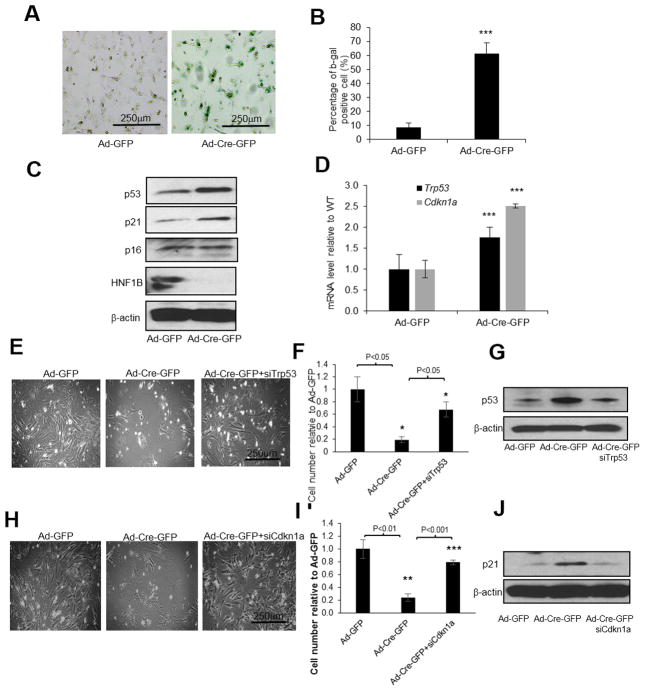
Figure 5. Inactivation of p53/p21 pathway rescued cell death induced by Hnf1b loss in MEFs.
Primary Hnf1b flox/flox P3 MEF cells were infected by Ad-Cre-GFP. After 48 h, b-gal senescence assay was performed (A) and quantification results are shown in (B). Protein levels of p53, p21, p16 as well as HNF1B were measured by immunoblotting (C). mRNA levels of Trp53 and Cdkn1a were determined by RT-PCR (D). Primary P3 MEFs were transiently transfected with either siRNA against Trp53 or Cdkn1a. After 24 h, cells were infected with Ad-GFP or Ad-Cre-GFP for an additional 48 h. Cell density images, quantification and corresponding protein levels by immunoblotting after Trp53 knockdown (E–G) or Cdkn1a knockdown (H–J) are shown. Data are expressed as mean±SD, N=3, *P<0.05; **P<0.01; ***P<0.001.

