Figure 1.
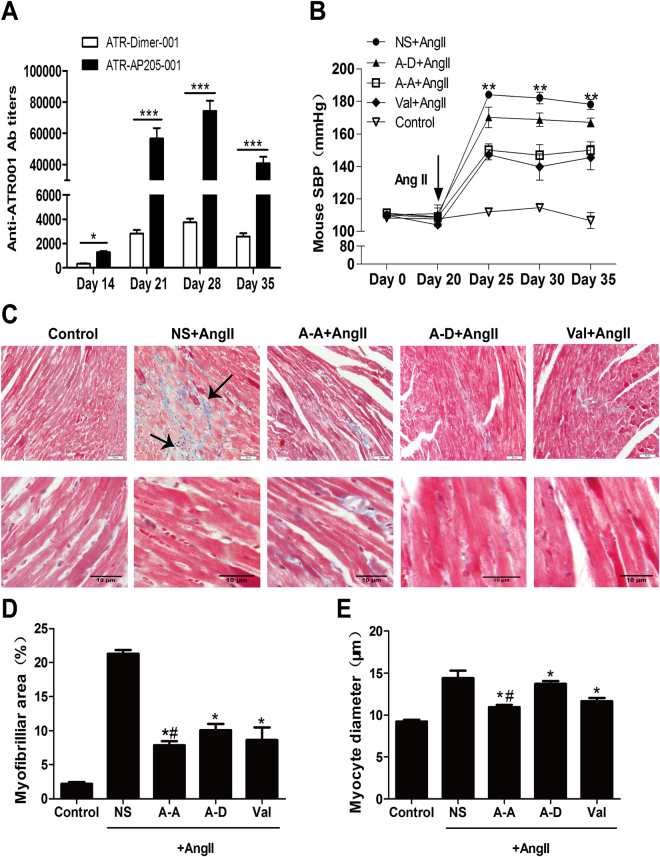
ATR-AP205-001 efficiently reduced blood pressure and target organ damages of Ang II induced hypertensive mice. (A) Anti-ATR001 antibody titers were measured by ELISA on days 14, 21, 28 and 35 after ATR-AP205-001 and ATR-Dimer-001 vaccination. n = 8 per group. *P < 0.05, ***P < 0.001 vs. ATR-Dimer-001. (B) Blood pressure of mice was tested by tail-cuff method on days 0, 20, 25, 30 and 35. **P < 0.01 vs. NS + Ang II group. (C) Masson trichrome staining identified fibrosis (marked by black arrows) of heart tissue in each group. Cardiomyocyte diameter was identified by the length perpendicular to the long axis of the cell (n = 20 myocytes per group). Scale bars, 50 μm (upper) and 10 μm (lower). (D) The ratio of fibrotic area to total heart tissue and (E) myocytes diameter tested in ImageJ. *P < 0.05 vs. NS + Ang II group, #P < 0.01 vs. ATR-Dimer-001 group. A-A: ATR-AP205-001, A-D: ATR-Dimer-001, Val: valsartan, and NS: natural saline in this Figure. Data are expressed as mean ± SEM.

