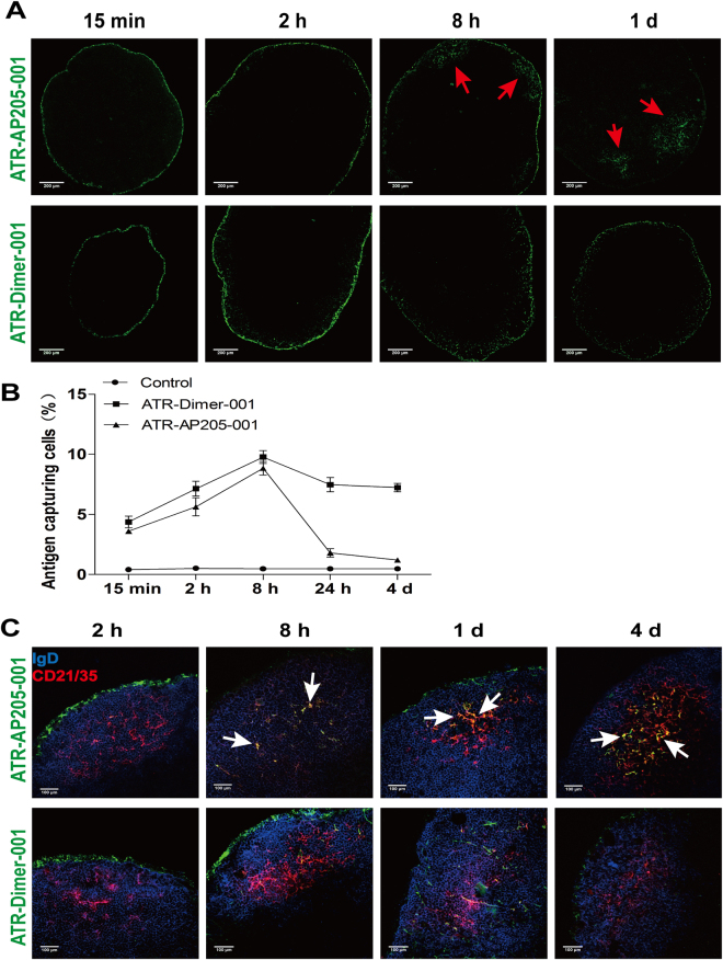
Figure 2.
ATR-AP205-001 was drained to popliteal lymph node and deposited in follicle dendritic cell (FDC) area. Mice popliteal lymph nodes were acquired at 15 mins, 2 hrs, 8 hrs, 24 hrs and 4 days after footpad injection. (A) Distribution of ATR-AP205-001 (upper, green) and ATR-Dimer-001 (lower, green) in popliteal lymph node. Red arrows figure out the accumulation of antigens inside the lymph node. Scale bars, 200 μm. (B) Proportion of cells participating in antigen-capturing. (C) Co-localization of ATR-AP205-001 (upper, green) and ATR-Dimer-001 (lower, green) with IgD+ follicle B cells (blue) and CD21/35+ FDCs (red). The antigens deposited in FDC area were annotated by white arrow. Scale bars, 100 μm. n = 6 per group. Data are expressed as mean ± SEM.

