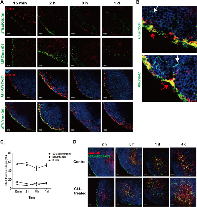
Figure 3.
Follicle B cells participated in ATR-AP205-001 transportation. (A) Co-localization of ATR -AP205-001 and ATR-Dimer-001 with DCs (CD11c+, red), SCS Mφs (CD169+, red) and follicle B cells (IgD+, blue) in dLNs. (B) Higher magnification co-localization images of ATR-AP205-001 and ATR-Dimer-001 with SCS Mφs and follicle B cells after 2 hours vaccination. Red arrows annotate vaccines co-localization with SCS Mφs, white arrows annotate vaccines carried by B cells. (C) Proportions of DCs (CD11c+), SCS Mφs (CD14+CD169+), and B cells (CD19+) gated on ATR001+ population after ATR-AP205-001 immunization. (D) Co-localization of ATR-AP205-001 with follicle B cells and FDCs in untreated and CLL-pretreated dLNs. n = 6 per group. Data are expressed as mean ± SEM. Scale bars, 100 μm.

