Figure 2.
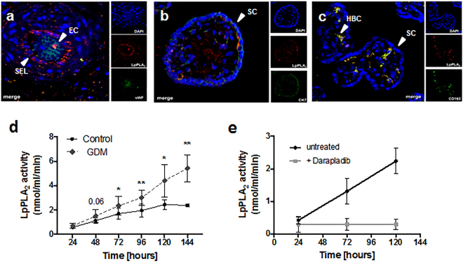
LpPLA2 is released by Hofbauer cells and increased in GDM. (a–c): Immunofluorescence staining of placental tissue. Images are representative of 3 independent experiments (N = 3). (a) van Willebrandt factor (vWF, green) was localized to the placental vessel lumen. LpPLA2 (red) was localized to villous stroma and sub-endothelial connective tissue layers. EC = endothelial cells, SEL = sub-endothelial layer. (b) Trophoblast marker Cytokeratin 7 (CK7, green) was present in the fused syncytial layer of the villous; LpPLA2 (red) was localized to villous stroma. SC = Syncytium. (c) Co-localization of LpPLA2 (red) with CD163 (green), a marker of Hofbauer cells, was observed within villous stroma. HBC = Hofbauer cells. (d) LpPLA2 activity secreted by HBCs isolated from healthy and GDM placenta (mean ± SD; N = 5 HBC isolations/group; two-way ANOVA). (e) LpPLA2 activity is abolished by addition of 150 nM Darapladib, a specific LpPLA2-inhibitor (mean ± SD, N = 4).

