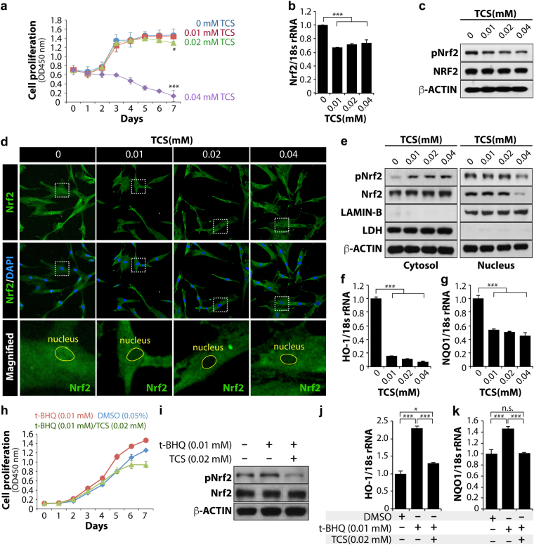
Figure 4.
TCS induces cytotoxicity in hMSCs by blocking the nuclear localization of Nrf2 protein. (a) Cell proliferation assay; each experiment was performed in triplicate. (b) EtOH or TCS-treated MSCs were collected at 24 hours to prepare cell lysates. The levels of Nrf2 mRNA were analysed by qRT-PCR. (c) The levels of total Nrf2 and phosphorylated Nrf2 proteins were determined by a western blot analysis. β-Actin was used as the loading control. (d) Immunofluorescence was performed to confirm the nuclear and cytosolic localization of Nrf2 proteins. DAPI was used to stain cell nuclei, and FITC-conjugated secondary antibody was used to visualize Nrf2 protein. The images were obtained using confocal microscopy. (e) Cytosolic and nuclear extracts were prepared and fractionated in accordance with the manufacturer’s instructions. The protein levels of total Nrf2 and phosphorylated Nrf2 were analysed by western blot in the EtOH and TCS-treated human cells. The protein level of LDH (lactate dehydrogenase) was used as a loading control for the cytosolic extracts and the protein level of LAMIN-B was used as a loading control for nuclear extracts. (f,g) HO-1 and NQO-1 mRNA levels were analysed by qRT-PCR. 18S rRNA was used as a loading control. (h–k) TCS blocks t-BHQ-induced Nrf2 nuclear localization and the expression of its target genes. (h) Cell proliferation assay. (i) The levels of total Nrf2 and phosphorylated Nrf2 proteins were determined by a western blot analysis. β-Actin was used as the loading control. (j,k) HO-1 and NQO-1 mRNA levels were analysed by qRT-PCR. 18S rRNA was used as a loading control. Standard deviation bars were calculated from at least three independent experiments. p < 0.05(*); p < 0.01(**); p < 0.001(***); Not statistically significant (n.s.).

