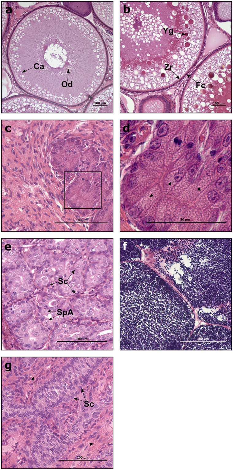
Figure 3.

Gonadal tissue from GCF and WT salmon. Histological images of Atlantic salmon ovaries (a–d) and testis (e–g). (a) Immature (oildrop stage) oocytes. (b) Early vitellogenic oocytes. (c) GCF ovary. (d) GCF ovary with lipid vacuoles (arrowheads), magnified from the marked area in (c). (e) Immature testis. (f) Mature testis containing mostly spermatozoa. (g) GCF testis with interstitial area (arrowheads). GCF, germ cell-free; Ca, cortical alveoli; Od, oil drop; Yg, yolk granule; Zr, zona radiata; Fc, follicle cell; SpA, spermatogonia A; Sc, Sertoli cell. Scale bar = 100 µm (a–c, e–g), 50 µm (d).
