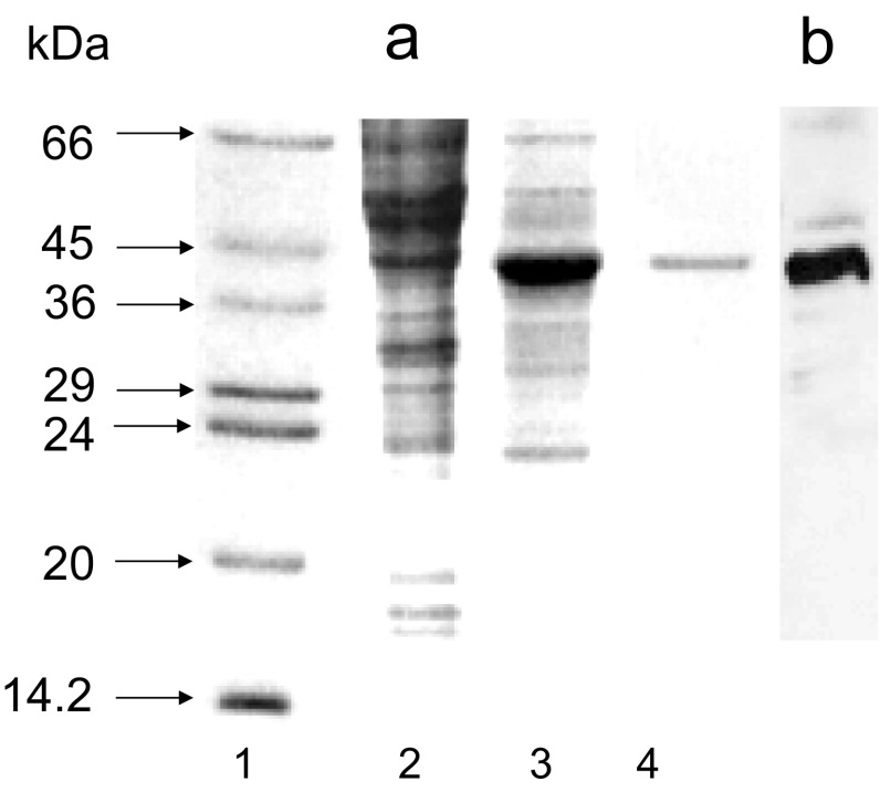
Fig. 1.
Expression of Nudt13 in High Five™ cells. High Five™ cells were infected with recombinant Nudt13 virus at a MOI of 10 for 48 h. Samples were analysed by SDS-PAGE (15% w/v) and stained with Coomassie Blue. a Lane 1 protein standards: bovine serum albumin (66 kDa), ovalbumin (45 kDa), glyceraldehyde 3-phosphate dehydrogenase (36 kDa), carbonic anhydrase (29 kDa), trypsinogen (24 kDa), soybean trypsin inhibitor (20 kDa) and α-lactalbumin (14.2 kDa); lane 2 control uninfected High Five™ cells; lane 3 high Five™ cells infected with Nudt13 virus for 48 h; lane 4 purified Nudt13. b Immunoblot analysis of Nudt13 (the same cells as in a, lane 3) using His.Tag monoclonal antibody

