Fig. 4.
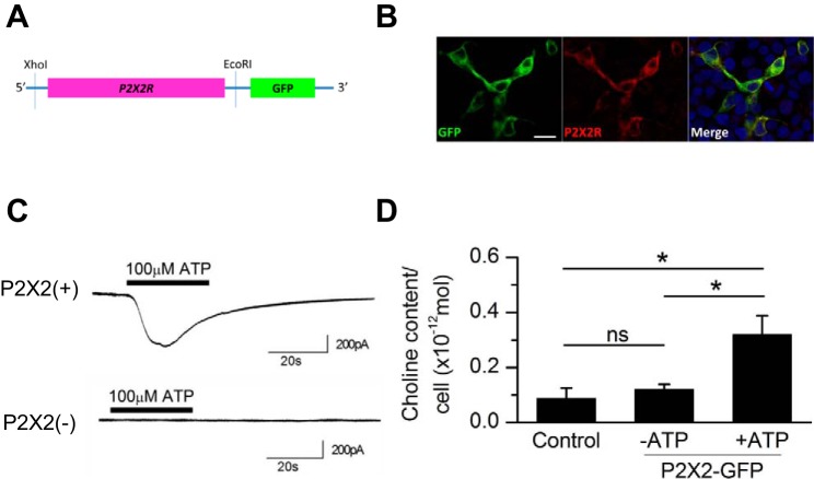
ATP-induced choline influx in transfected HEK-293T cells expressing the fusion protein of P2X2 purinoceptors and GFP. A: design of the cDNA of P2X2 purinoceptors fused with GFP. B: immunoreactivity for P2X2 purinoceptors in HEK-293T cells transfected with the P2X2R::GFP vector. Bar, 20 μm. Green, GFP; red, P2X2 purinoceptors; blue, DAPI. C: responses to ATP in GFP-positive cells [P2X2(+)] and GFP-negative cells [P2X2(−)] in choline-rich (Ca-free) solution (no. 2-3). Horizontal bars above the traces show the timing of ATP application. The intracellular solution was KCl (HEK) (no. In-3). D: effects of ATP on the cytosolic choline content in HEK-293T cells. Experiments were conducted in choline-rich solution (no. 2-2). Control cells were collected from the well without transfection; P2X2-GFP cells were collected from the well with transfection with (+ATP) or without 100 μM ATP (−ATP). Values are means ± SE; n = 4 for all 3 experimental conditions. *P < 0.05; n.s., not significant.

