Fig. 1.
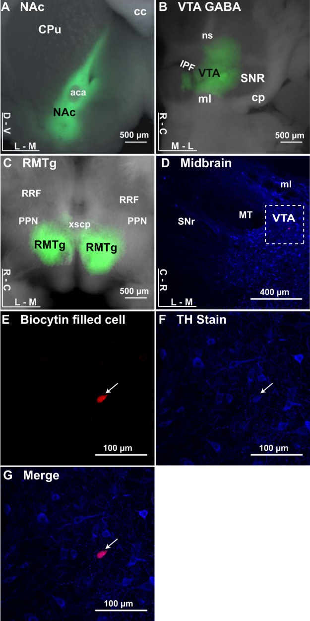
ChR2 expressed at GABA inputs to dopamine neurons. A: image of a coronal brain section expressing ChR2 in the nucleus accumbens (NAc; green). aca, Anterior commissure; cc, corpus callosum; CPu, caudate/putamen; D, dorsal; V, ventral; L, lateral; M, medial. B: image of a horizontal brain section expressing ChR2 in the ventral tegmental area (VTA; green). cp, Cerebral peduncle; IPF, interpeduncular fossa; ml, medial lemniscus; ns, nigrostriatal bundle; SNR, substantia nigra pars reticulata; R, rostral; C, caudal. Local VTA GABA neurons were targeted in Vgat-ires-Cre mice using injections of the rAAV5/EF1α-DIO-hChR2-(H134R)-mCherry viral construct. C: image of a horizontal brain section expressing ChR2 in the rostromedial tegmental nucleus (RMTg). RRF, retrorubral field; PPN, pedunculopontine nucleus; xscp, decussation of the superior cerebellar peduncle. D–G: filled neuron costained with tyrosine hydroxylase (arrows). D: horizontal midbrain section stained for tyrosine hydroxylase (TH; blue) with a recorded neuron (biocytin fill; red), with dashed inset reference for filled neuron in E–G. E: VTA neuron filled with biocytin during recording, labeled with streptavidin coupled to Alexa594. F: TH staining of slice with rabbit anti-TH primary and goat anti-rabbit secondary. G: merged image showing biocytin-filled neuron is costained with TH.

