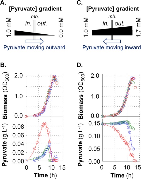
FIG 3 .
Functional characterization of the PftAB pyruvate transporter in B. subtilis. (A and C) Schematic representation of a facilitated transport of pyruvate in the absence (A) or presence (C) of extracellular pyruvate. mb., membrane. (B and D) Growth of the B. subtilis WT (blue), ΔpftAB (green), and ΔpftAB PhspftAB (red) strains and corresponding concentrations of extracellular pyruvate. The results from a representative experiment are shown. (B) Cells were grown in M9G with 200 µM IPTG. (D) Cells were grown in M9G with 200 µM IPTG and 0.15 g ⋅ liter−1 pyruvate.

