
Figure 1.
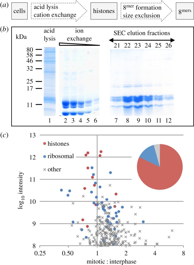
Purification of histones from asynchronous and nocodazole arrested cells. (a) A scheme for purification of histones from DT40 cells while preserving cell-cycle-regulated post-translational modifications. (b) A typical Coomassie-stained SDS-PAGE gel of purification steps: Lane 1—acid lysis supernatant. Lanes 2–6—dilution series of pooled ion-exchange eluate. Lanes 7–12—elution fractions after size exclusion chromatography (SEC). (c) Summary of mass spectrometry (MS) analysis of the isolated histones. Plot of log10 of peptide intensities and log2 of mitotic enrichment. Red, histones; blue, ribosomal proteins; grey, other contaminating proteins. Inset pie chart shows the total sum of peptide intensities for these proteins.