Figure 2.
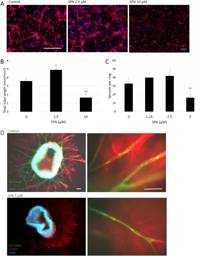
Effect of SFN on tube formation of HUVECs and microvessel sprouting in mouse aortic rings. (A) Representative pictures from 3D co-culture of HUVECs and pericytes M2 at day 4 with SFN 0, 2.5, 10 µM. HUVEC were identified by immunodetection of CD31 (red), nuclei were stained (blue) and merged pictures are shown. Scale bar = 500 µm. (B) Total lengths of CD31 positive tubes were measured and expressed as mean ± SD (n ≥ 5), *p < 0.05, **p < 0.01 compared to control. (C) Dose response of SFN on microvessel sprouting from aortic rings embedded in collagen with DMSO (0.1%) as control. Data are presented as mean ± SD (n ≥ 5), **p < 0.01 compared to control. (D) Representative pictures from the immunofluorescent staining of aortic rings. Endothelial sprouts were stained with BS1-lectin-FITC (green), supporting cells were stained for α-smooth muscle actin (red), nuclei were stained with DAPI (blue) and merged pictures are shown. Scale bar = 100 µm.

