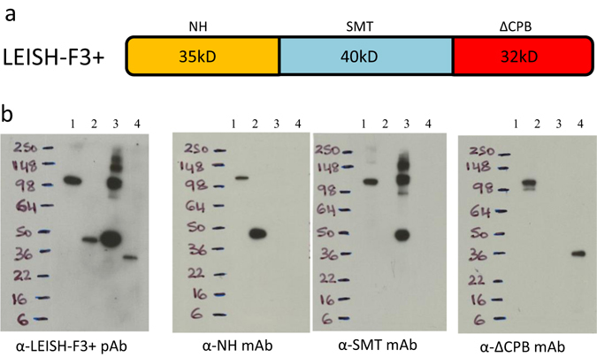
Fig. 2.

Construction and characterization of the LEISH-F3+ fusion protein. In a, a cartoon depiction of the LEISH-F3+ fusion protein is shown. In b, LEISH-F3+ fusion protein was characterized by immunoblot. Recombinant LEISH-F3+ (lane 1), Nucleoside hydrolase (NH; lane 2), Sterol-24-c-methyl-transferase (SMT; lane 3) or truncated cysteine protease B protein (ΔCPB; lane 4); were loaded at 100 ng each lane into gels. Blots were developed with mouse polyclonal or monoclonal antibodies as indicated, derived from the same original gel. The cartoon depiction of the fusion protein is the authors own rendering
