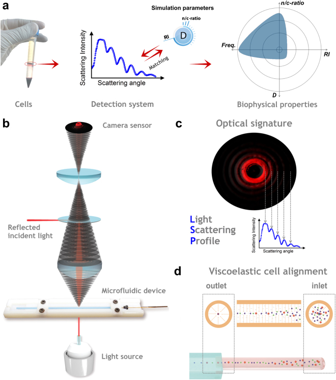
Figure 1.
Schematic design of the detection procedure. (a) Working principle of the analysis of multiple biophysical properties of a cell, with the following steps: cell collection via density gradient separation; LSP matching with simulation parameters (LSP is calculated from the optical signature); radar plot of biophysical properties from the investigated cells (properties are defined in Fig. 2b, ‘Freq.’ indicates the relative cell frequency). (b) Schematic design of the detection system. The incident light source passes the microfluidic alignment stage from the bottom, while the scattered light collection stage maps the obtained optical signature of each passing cell on the sensor (not shown for easier readability). The incident light is reflected out of the signature by a small beam stopper, placed between the optical lenses. (c) LSP is calculated out of the signature by averaging the intensity values of all the sensor pixels with same scattering angle. (d) The viscoelastic cell alignment over distance in a round capillary is illustrated (flow direction is from the right to the left).

