Fig. 1.
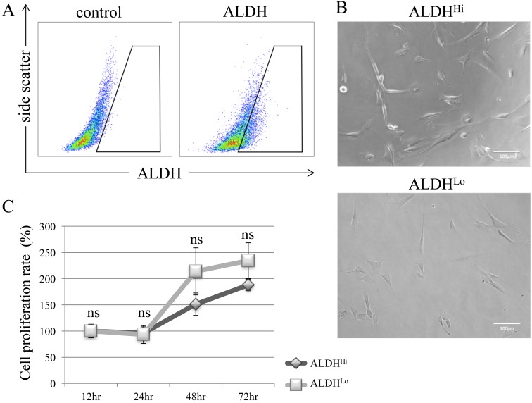
Detection of ALDH-positive subpopulations of canine ADSCs and evaluation of proliferation rates. (A) Flow cytometric analysis of canine ADSCs. Baseline fluorescence was established by adding the ALDH inhibitor diethylaminobenzaldehyde (control). (B) Sorted ALDHHi and ALDHLo showed similar morphologies after incubation for 24 hr. (C) There was no significant difference in cell proliferation rates between ALDHHi and ALDHLo subpopulations. Values are expressed as mean ± standard error (n=5). *P<0.05 vs. control cells; ns, not significant.

