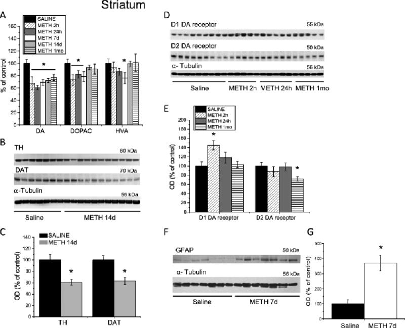
Fig. 2.

Changes in the markers of DA system integrity and GFAP in the dorsal striatum following METH self-administration. Rats self-administered METH for eight 15-h sessions and were euthanized at 2 h, 24 h, 7 days, 14 days or 1 month withdrawal. (A) METH caused significant decreases in striatal DA levels up to 1 month post-drug. DOPAC concentrations were decreased at 2 h, 24 h, and 7 days, but returned to control levels after 14 days of withdrawal. HVA levels were transiently decreased at 7 days after cessation of METH self-administration, but normalized at 14 days post-drug. (B) Representative immunoblots showing TH and DAT protein levels in the striatum at 14 days after METH withdrawal. (C) Quantitative analyses demonstrated reductions in DAT and TH protein levels in the striatum of METH-treated rats. (D) Representative immunoblots of D1 and D2 DA receptor protein levels. (E) METH self-administration caused increases in D1 DA receptor protein levels at 2 hours post-drug; D2 DA receptor protein levels were decreased after 1 month withdrawal. (F) A representative immunoblot demonstrating GFAP levels in rat striatum 7 days following cessation of METH self-administration. (G) Quantitative analyses of the Western blots show increases in GFAP levels in the METH-treated rats. N = 7–11 per group. Data are expressed as percent (protein optical density, monoamine levels) of mean ± SEM values of saline group. Data were analyzed by ANOVA followed by PLSD. * p < 0.05, significantly different from saline group. Figure adapted from Krasnova et al. 2010, 2013.
