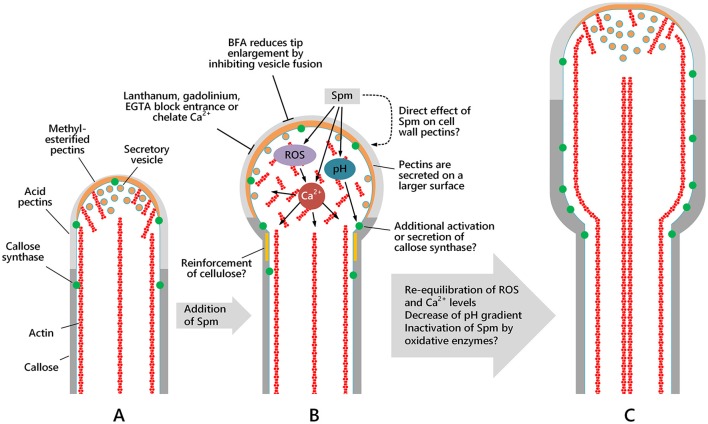
Figure 13.
Schematic drawing showing the morphology of the apical region and the spatial distribution of actin filaments, secretory vesicles and cell wall components in pollen tubes and how Spm perturbs either their distribution or the morphology of the apical region. The pollen tube sub-apex is characterized by the actin fringe while in the shank region actin filaments form regular and longitudinal cables, which are essential for organelle and vesicle movement. Spm profoundly alters Ca2+, H+, and ROS concentrations and distribution, thereby affecting not only microfilament organization but also vesicle delivery. As a final result, assembly of the cell wall and shaping of the growing pollen tube tip is altered. (A) Pollen tube after 1 h of germination; (B) pollen tube after 1 h of germination in standard medium, then supplied with Spm for one additional hour; (C) pollen tube grown for 1 h in standard medium and then supplemented with Spm for two additional hours. As yet unknown mechanisms re-equilibrate ion distribution and concentration and allow the pollen tube to resume growth even though tube diameter remains larger and growth rate slower. Inactivation of Spm by oxidative enzymes cannot be excluded.

