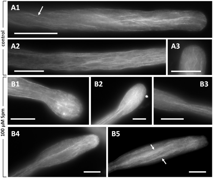
Figure 5.
Distribution of actin filaments in pear pollen tubes. (A1,A2) Control pollen tubes. The arrow indicates a longitudinal helicoidally-arranged actin filament. (A3) Detail of a control pollen tube apex. (B1–B5) Pollen tubes treated with Spm. (B1) A balloon-shaped pollen tube showing completely disorganized actin filaments in the apex. (B2) Reorganization of actin filaments at the snake-shaped stage. (B3) When the pollen tube re-starts to grow, actin filaments run along the cortical region and open like a fan at the neck level. (B4,B5) Actin filaments can be observed as the new shovel-shaped pollen tube resumes growth. Arrows indicate some actin bundles that run longitudinally along the middle and the cortex of pollen tubes. Bars = 10 μm. Data are representative of three independent experiments.

