Figure 6.
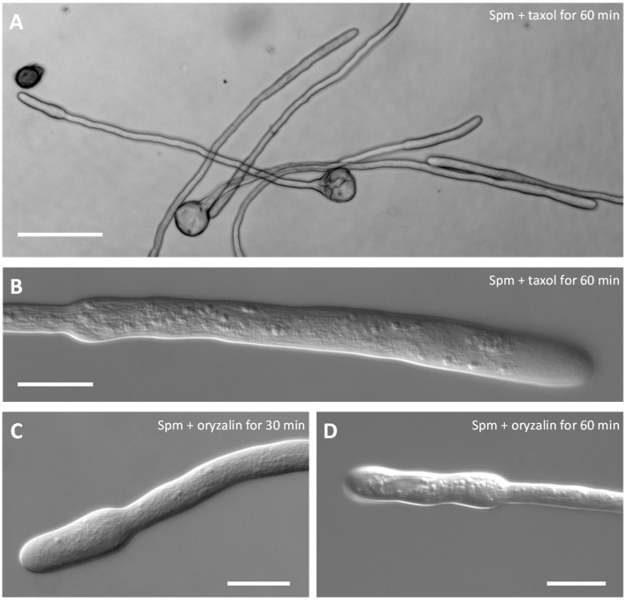
Co-treatment of pear pollen tubes with Spm and microtubule inhibitors. (A) DIC view of pollen tubes after co-treatment with Spm and taxol. (B) Detail of a single pollen tube after Spm+taxol treatment (DIC view). (C,D) DIC views of pollen tubes after co-treatment with Spm and oryzalin. Neither inhibitor affected the formation of the shovel shape. Images were captured after the incubation time indicated in each picture. Both analyses were performed at least until 1 h after supplementation of Spm and drugs. Bar in (A) 100 μm; bars in (B–D) 20 μm.

