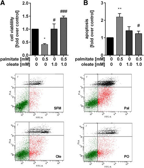
Fig. 1.

Palmitate-induced cell death is rescued by oleate. HepG2 cells were stimulated with palmitate (0.5 mM), oleate (1 mM) and a combination of both (0.5 mM/1 mM) for 48 h. a Cell viability was measured by WST-1 assay. b Apoptosis was determined by flow cytometrical counting of Annexin V-FITC and Annexin V-FITC/PI positive stained cells. Data were normalised to control (serum free medium) which was set 1. Representative dot plots of the AnnexinV-FITC/PI staining in HepG2 cells are shown. Data represent three independent experiments performed in triplicates shown as means ± SEM. *p < 0.05, **p < 0.01 compared to control. # p < 0.05, ### p < 0.001 compared to palmitate. SFM: serum free medium; Pal: palmitate; Ole: oleate; PO: palmitate/oleate
