Fig. 1.

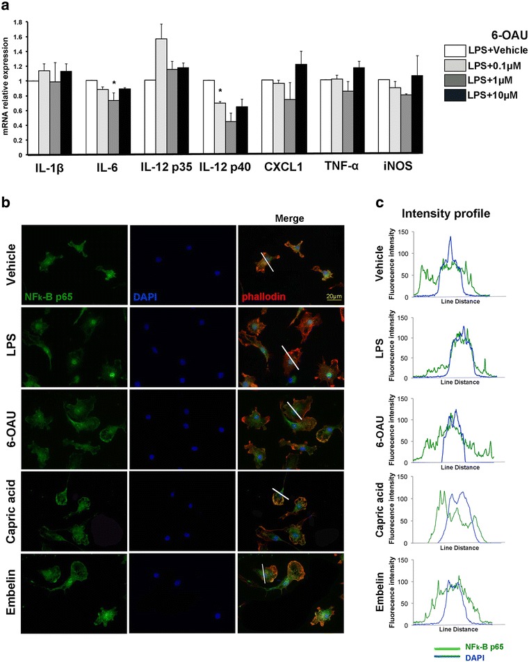
GPR84 does not modulate pro-inflammatory responses in cultured microglia. a Primary cultured microglia were treated with bacterial lipopolysaccharide (LPS) and either vehicle or 6-OAU for 16 h. mRNA for interleukin (IL)-1β, IL-6, IL-12 p35, IL-12 p40, chemokine (C-X-C motif) ligand 1 (CXCL1), tumor necrosis factor (TNF)-α, and inducible nitric oxide synthase (iNOS) was quantified by quantitative real-time RT-PCR. Results were normalized to LPS-treated samples. Values represent mean ± SEM from three independent experiments. *p < 0.05. b Microglia were treated with vehicle, 100 ng/mL LPS, 1 μM 6-OAU, 1 μM capric acid, and 1 μM embelin for 16 h. Cells were stained with anti-nuclear factor kappa B (NF-κB) p65 antibody (green), Alexa Fluor 594-labeled phalloidin (red), and DAPI (blue). c Line scan graphs of representative cells show the fluorescent intensity of p65 (green) and DAPI (blue) along white lines. Scale bar, 20 μm
