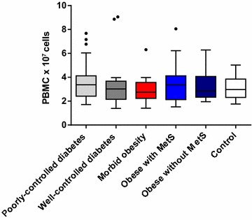
Fig. 2.

Mean PBMC cell count (× 107 cells): both visits (Visit 1 and Visit 2) combined. PBMC peripheral blood mononuclear cell. The bottom and top edges of the box are located at the sample 25th and 75th percentiles and the center horizontal line is drawn at the 50th percentile (median). The whiskers extend at most 1.5 interquartile ranges
