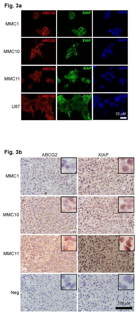
Fig. 3. ABCG2 and XIAP expression in matching patient tumors and tumorsphere lines.
a) Immunofluoresence analyses of patient-derived tumorsphere lines MMC1, MMC10 and MMC11 and U87 cells stained with antibodies against ABCG2 (red), and XIAP (green). All nuclei are stained with DAPI (blue). b) immunohistochemistry (IHC) images of MMC1, MMC10, and MMC11 patient tumors using antibodies against ABCG2 and XIAP. IHC scores for MMC1, MMC10 and MMC11 were 40, 160 and 180 respectively for ABCG2, and 100, 140 and 100 respectively for XIAP.

