
Figure 5.
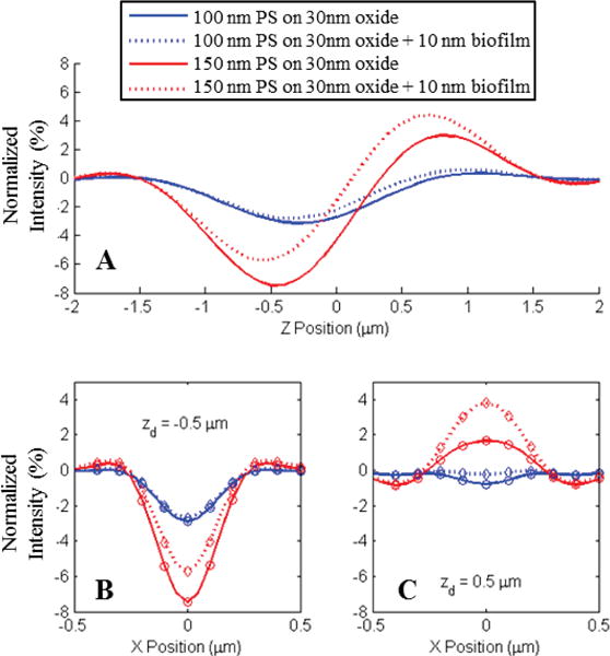
A) Shift in the normalized intensity of center pixels for two sizes of polystyrene spheres (100nm and 150nm) due to the presence of a 10 nm biofilm of capture probes. B–C) Line profiles of the observed appearance for the three nanospheres simulated for two focal plane offsets (−.5 and +.5 μm) with respect to the oxide surface. Simulations were generated for an .9NA in-water imaging system. Circles in plots B and C represent pixel sampling locations for a 2 μm pixel pitch with 40× magnification.