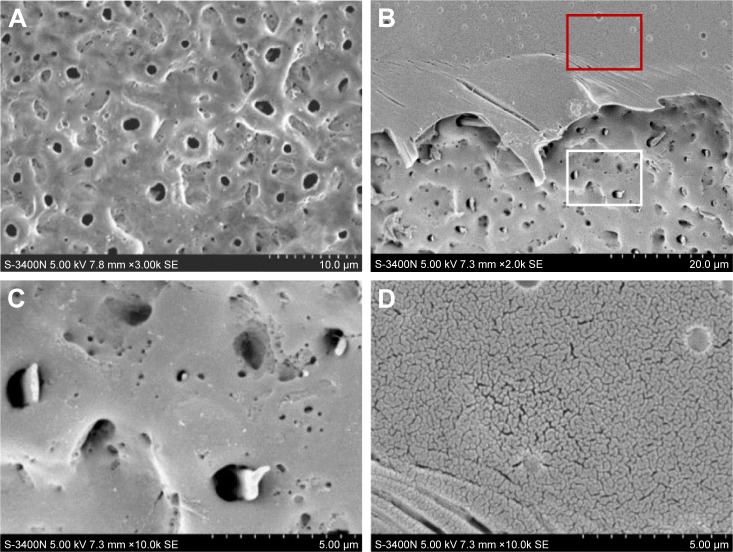
Figure 2.
Surface morphology of original MAO titanium implant and MAO titanium implant with PLGA coating. Representative SEM image of the original MAO titanium implant surface (A). The border between PLGA coating and titanium implant (B). High magnification image of the white rectangle area in B showing the PLGA penetrated into the micropores of MAO implant (C). High magnification image of the red rectangle area in B displaying the surface of PLGA coating (D).
Abbreviations: SEM, scanning electron microscopy; MAO, microarc oxidation; PLGA, poly(lactic-co-glycolic acid).

