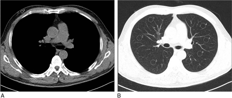
Figure 2.

An example of ROI of measurements on transverse unenhanced multidetector CT images. For all measurements, the size, shape, and position of the ROIs were kept approximately constant across patients. (A) ROIs manually drawn on the descending aorta (ROI 1), pulmonary trunk (ROI 2), subcutaneous fat of the anterior abdominal wall (ROI 3), paraspinal muscle (ROI 4) and air (ROI 5); (B) ROIs manually drawn on the pulmonary parenchyma (ROIs 1–6). CT = computed tomography, ROI = range of interest.
