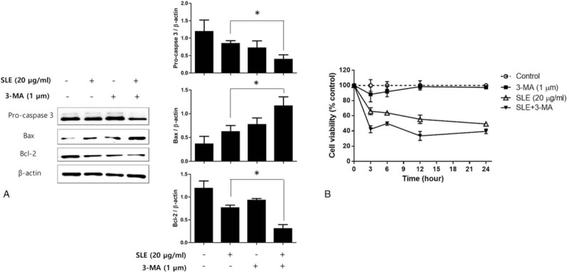
Figure 5.

Effect of autophagy inhibition on apoptosis in SLE-treated LNCaP cells. LNCaP cells were cotreated with 1 μM 3-MA and 20 μg/mL SLE, and (A) the expression of caspase 3, Bcl-2, and Bax was measured using western blot analysis, and (B) the cell viability was determined by MTT assay at 3, 6, 12, and 24 h of treatment. β-actin was used as a loading control. Expression of Pro-caspase 3, Bax, and Bcl-2 was quantified by scanning densitometry. The relative intensities were expressed as the ratio of Pro-caspase 3, Bax, and Bcl-2 to β-actin. Representative data from 3 independent experiments are shown and quantitated. Values are mean ± S.E.M of 3 independent experiments. 3-MA = 3-methyladenine. ∗P < .05, significantly different from the control group. LNCaP = lymph node carcinoma of the prostate, MTT = 3-(4,5-dimethylthiazol-2-yl)-2,5-diphenyltetrazolium bromide.
