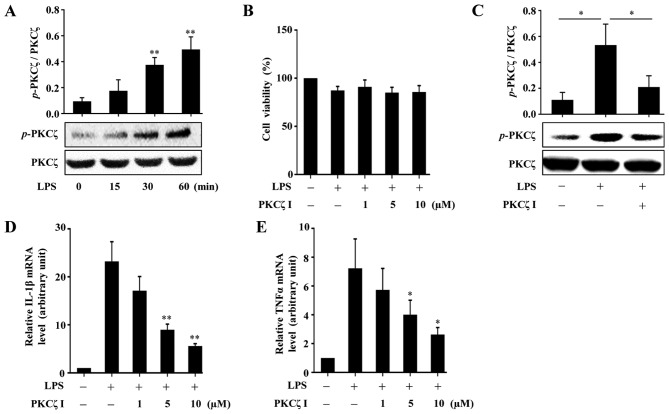
Figure 1.
The roles of protein kinase Cζ (PKCζ) in lipopolysaccharide (LPS)-induced inflammatory responses in RAW264.7 macrophages. (A) Representative immunoblots assessing the changes in the levels of phosphorylated PKCζ in RAW264.7 cells following treatment with 100 ng/ml LPS at indicated time points (0, 15, 30 and 60 min). (B) CCK-8 assay assessing the effects of LPS (100 ng/ml) alone or with PKCζ specific pseudosubstrate inhibitor (PKCζ I) (1, 5 or 10 µM) on the viability of RAW264.7 cells. Data represent means ± SD of 3 independent experiments, the value in LPS-free group arbitrarily set as 100%. (C) Representative immunoblots showing the effects of PKCζ I (10 µM) on LPS-induced PKCζ phosphorylation. (D and E) RT-qPCR analysis showing the effects of PKCζ I on LPS-induced mRNA level changes of interleukin-1β (IL-1β) or tumor necrosis factor α (TNFα). Each bar represent the means ± SD of 3 independent experiments, and normalized to corresponding total PKCζ protein level (A and C) or GAPDH mRNA level with the value in the LPS-free group set as 1 arbitrarily (D and E); *p<0.05 and **p<0.01 vs. the value at 0 min (A) or the value in corresponding LPS alone group (C–E).

