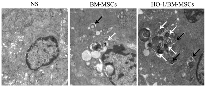
Figure 4.
Ultrastructure and autophagic vacuoles of the transplanted liver on POD 7 observed under a transmission electron microscope (magnification, ×25,000). The normal saline (NS)-treated group presented nuclear pycnosis, an irregular nuclear membrane with deformation, significantly swollen endoplasmic reticulum and mitochondria and no typical autophagic vacuoles. The BM-MSCs-treated group showed slight nuclear pycnosis and deformation, no obvious edema of the endoplasmic reticulum, slight edema of the mitochondria, and visible initial and degraded autophagic vacuoles. The HO-1/BM-MSCs-treated group reported slight nuclear pycnosis without deformation, no edema in endoplasmic reticulum or mitochondria, and a large number of initial autophagic vesicles and degraded autophagic vesicles. The black arrow shows initial autophagic vesicles, containing complete rough endoplasmic reticulum and mitochondria. The white arrow shows degraded autophagic vesicles, containing degraded rough endoplasmic reticulum and mitochondria. POD, postoperative day; BM-MSCs, bone marrow mesenchymal stem cells; HO-1/BM-MSCs, HO-1 transduced BM-MSCs.

