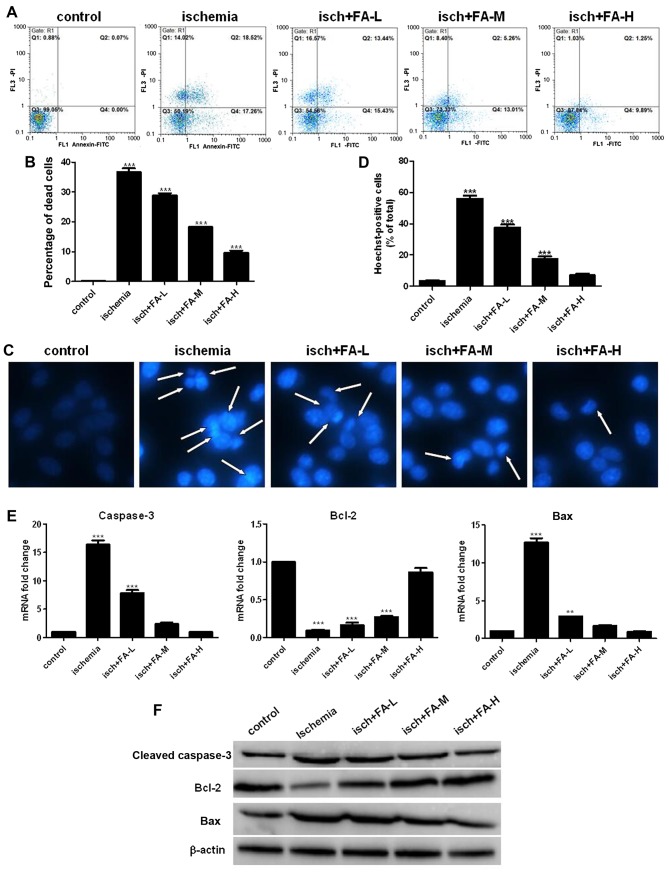
Figure 5.
Effect of ferulic acid (FA) on thye ischemia-induced apoptosis of PC-12 cells. (A) Annexin V/PI staining was used to determine apoptosis by flow cytometry. (B) Histograms showing the ratio of dead cells to the total nuclei. (C) Nuclearmorphology was determined by Hoechst 33258 staining; the white arrows indicate the apoptotic cells, at ×400 magnification. (D) Histograms showing the ratio of condensed nuclei to total nuclei. (E and F) The expression of apoptosis related proteins was examined by qPCR and western blot analysis.

