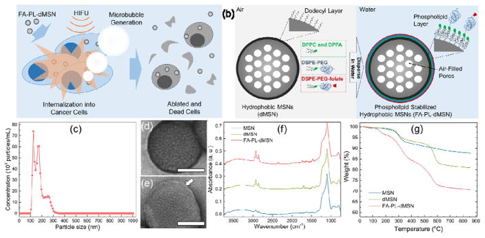
Figure 1.
Design and characterization of nanoscale ultrasound agents. a) Schematic representation of microbubble generation and cancer cell elimination by phospholipid coated mesoporous silica nanoparticles upon HIFU exposure. b) Schematic representation of phospholipid coated mesoporous silica nanoparticle preparation. c) Size distribution of FA-PL-dMSN in PBS (pH 7.4, 10 mM) as determined by Nanoparticle Tracking Analysis. TEM images of d) MSN and e) FA-PL-dMSN with uranyl acetate stain. Arrow in e) indicate the phospholipid layer around the MSNs. f) FTIR spectra of MSN, dMSN, and FA-PL-dMSN. g) TGA spectra of MSN, dMSN, and FA-PL-dMSN. Scale bars in (d) and (e) are 50 nm.

