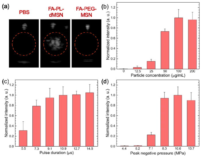
Figure 2.
Microbubble generation by nanoscale ultrasound agents upon HIFU exposure. a) Representative images were taken from movies acquired during HIFU irradiation in the presence or absence of nanoparticles (200 μg mL−1). Microbubble generation was only observed in the presence of FA-PL-dMSN. b) Normalized ultrasound contrast intensities from the acquired movies of FA-PL-dMSN samples at different concentrations were exposed to HIFU at a pulse duration of 10.9 μs and peak negative pressure of 10.6 MPa. c) Normalized ultrasound contrast intensities from the acquired movies of FA-PL-dMSN samples (100 μg mL−1) were exposed HIFU with different pulse durations at a peak negative pressure of 10.6 MPa. d) Normalized ultrasound contrast intensities from the acquired movies of FA-PL-dMSN samples (100 μg mL−1) were exposed to HIFU with different peak negative pressures at pulse duration of 10.9 μs. Error bars = 1 SD, studies were run in triplicate.

