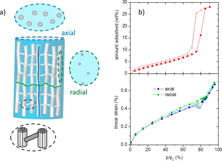
Figure 3.
(a) Schematic of the microstructure of a sheared sample of cylindrical shape; the dashed circles represent cross sections in two different directions (blue and green) and a magnification (black). (b) Adsorption (top) and strain isotherms (bottom) of the sheared sample during H2O adsorption at 17 °C. Full symbols denote adsorption, open symbols denote desorption. The strain isotherms have been measured on cubic samples (cut from the cylindrical monoliths) in the two different directions.

