
Figure 4.

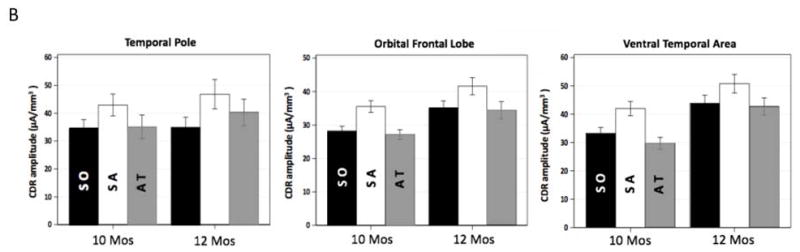
Development of the effect of sustained attention on infant theta source activation. A. 3D displays for the difference in CDR amplitude between sustained attention and attention termination separately for the four ages. Age-appropriate average MRI templates were used for the display for each age. Sustained attention effect was primarily shown in the temporal pole, orbital frontal, and ventral temporal regions, especially for 10 and 12 months. B. Bar graphs for the average CDR amplitudes in these brain networks for 10 and 12 months. Error bars represent SEMs.