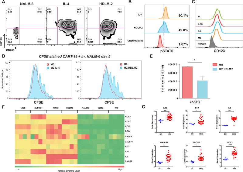
Figure 2. HL cells polarize normal macrophages to a M2-like phenotype.
A. Human normal donor macrophages differentiated from peripheral blood monocytes were cultured with a control acute lymphoblastic leukemia cell line (NALM-6), IL-4 (M2 positive control), or HL cell line HDLM-2 conditioned supernatant. HDLM-2 cells can polarize macrophages toward an M2 phenotype (CD163+CD206+) after a 24-hour culture. B. HDLM2 supernatants trigger phosphorylation of STAT6 on macrophages, similarly to IL-4. C. M0, M2-polarized (IL-4 or IL-13), and HL polarized macrophages express CD123 by flow cytometry. D. M2-polarized (IL-4) and, to a greater extent, HL-polarized macrophages can reduce anti-CD19 chimeric antigen receptor T cell proliferation in response to irradiated CD19+ NALM-6 target cells, as shown by CFSE (Carboxyfluorescein succinimidyl ester - a fluorescent cell stained dye that is diluted with cell proliferation) dilution assay. E. HL-polarized macrophages reduce CART19 proliferation in response to irradiated NALM-6 target cells, as shown by absolute T cell numbers at day 5 F. Heatmap demonstrating that HL cell lines HDLM2, KMH2, SUPHD1, and L428 secreted significantly more myeloid cell recruiting chemokines (CCL2, CCL3, CCL4, CCL5, CXCL9, CXCL10), M2 polarizing cytokines (IL13, IL10, IL6) and macrophage supporting factor GM-CSF than control non-HL cell lines K562 and NALM6 or media alone. G. RNA expression analysis of 29 microdissected HRS cells (dataset GSE-39133) confirmed the expression of M2 polarizing cytokines (IL13, IL10, IL6), macrophage supporting factors (GM-CSF, M-CSF), and the immunosuppressive checkpoint ligand PDL1 in primary samples.

