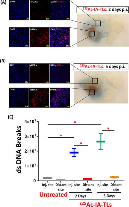
Figure 6. Double strand DNA breaks present within tumor tissue but absent within surrounding tissue showing enhanced vascular permeability.

Immunohistochemical staining of sectioned mouse brains revealed the presence of γH2A.X within tumor tissue, indicating α-particle induced tumor cell killing 2 days (n=3) (A) and 5 days (n=3) (B) post intracranial infusion (p.i.) of 225Ac-IA-TLs. (C) Immunohistochemical staining of sectioned mouse brains also revealed negligible presence of γH2A.X in normal brain tissue surrounding tumors (0.5mm from GBM border) (n=3), indicating absence of double strand (ds) DNA breaks within these regions where enhancement in vascular permeability (extravasation of Evans Blue dye) was observed. Data represented as mean +/− SEM. Student’s t-test and One-way ANOVA was performed to assess difference between experimental groups (*p< 0.001(significant)).
