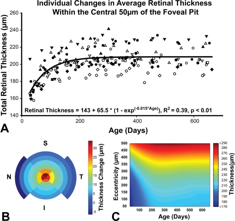
Figure 6.

A. The average retinal thickness within 50μm of the pit increased by 20.9% with the majority of change occurring within the first 150 days after birth. The trend was similar for all animals and was best fit with an exponential to maximum function. Each subject is represented by a different symbol. B. The figure illustrates total retinal thickness change for each quadrant at the eccentricities illustrated in figure 3E. C. A surface plot illustrating changes in average thickness as a function of age and eccentricity up to 500 μm.
