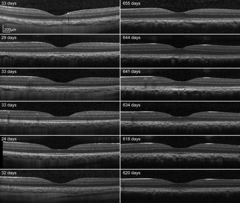
Figure 7.

Scaled SD OCT images for six subjects at baseline (left) and endpoint (right) through the fovea center. A well defined foveal pit is seen in all animals at the first scan session. The overall shape of the foveal pit does not change, but there is an increase in outer retinal thickness on the endpoint scans.
