Figure 2.
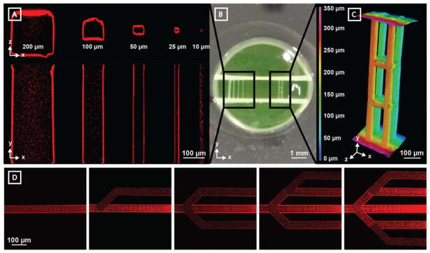
Microvessel generation is readily controlled in 3D with micron-scale resolution. Vertical channel sets were generated between two horizontal channels (~500 μm diameter) spaced ~1.5 mm apart. Vessel perfusion with fluorescent polymer beads (red, 0.22 μm diameter) enabled channel visualization by multiphoton microscopy. A) Channels with a wide range of cross-sectional sizes (200 μm x 200 μm, 100 μm x 100 μm, 50 μm x 50 μm, 25 μm x 25 μm, and 10 μm x 10 μm) are fully patent. B) Photographic image of hydrogel displays both parallel microchannel and 3D multilayered channel sets. C) Photodegraded channels can be generated with full 3D control. Four parallel 50 μm x 50 μm (width x height) channels generated in the Y direction (X-spacing = 150 μm, Z-spacing = 100 μm) connect the horizontal channels. 8 bridging channels (50 μm x 50 μm, cross sectional) interconnect features in the X and Z directions. Microbead fluorescence is viewed using Z-depth color-coding. D) Perfusable vascular network geometry can be altered over time through iterative network photodegradation (channel cross section of 40 μm x 40 μm, red beads = 0.22 μm diameter).

