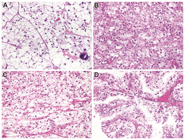
Figure 1.
Morphology of Genetically Confirmed Xp11 translocation RCC in this study. Panel A: This tumor has clear cell features and psammoma bodies; Panel B: This tumor closely resembled clear cell RCC; Panel C: This primary tumor resembled clear cell RCC; and Panel D: Recurrence of the primary tumor shown in C demonstrates papillary architecture. All images are taken at 400X magnification, and all are Hematoxylin and Eosin stained.

