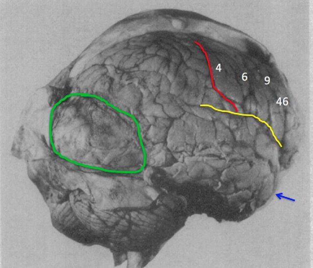
Figure 1.
The brain of Patient A following “bilateral” frontal lobectomy (data from Brickner, 1952). Patient A was reported as the first human subject to undergo “bilateral” frontal lobectomy. Although this patient has previously been described as a classic example of how lesions to PFC do not impair consciousness (Koch et al., 2016a), simple visual inspection of this patient's postmortem brain image reveals that massive amounts of residual right PFC remained following the surgery. Therefore, the fact that this patient exhibited signs of consciousness following surgery is not surprising. This patient also had a large posterior meningioma exerting pressure on extrastriate cortices. The green region marks the posterior meningioma, the red line labels the central sulcus, the yellow line shows the Sylvian fissure, and the blue arrow marks the temporal tip. Brodmann areas are marked by white numbers.

