Figure 5.
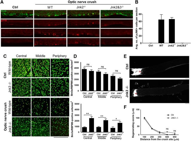
JNK3 knock-out promoted RGC survival but not axon regeneration. A, Immunofluorescence for retinal ganglion cells using β-III tubulin (green) and phosphorylated JNK (red) in wild-type control retinal sections and after optic nerve injury in wild-type mice, Jnk2−/−, and Jnk2 and 3−/−. B, pJNK quantifications for control and optic nerve injury. C, Images of the retina showing RGC-specific markers RBPMS (green) and Brn3a (yellow) in JNK3 knock-out (KO) and age-matched wild-type (WT) mice before and 2 weeks after optic nerve crush. D, RGC quantification demonstrated similar RGC density in JNK3 KO and WT control mice before the optic nerve injury. Two weeks after optic nerve injury, however, RGC density in JNK3 KO mice was significantly higher than in WT animals (*p < 0.05, **p < 0.001, one-way ANOVA; n = 5 per group; NS, Not significant). E, Images of the optic nerve sections showing CTB-labeled axons 2 weeks after optic nerve injury. Asterisks indicate lesion sites. F, A similar number of regenerating fibers was observed in JNK3 KO and WT control animals. Error bars indicate SE. Scale bar, 200 μm.

