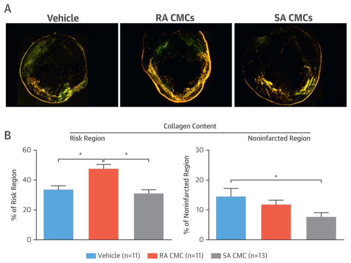
FIGURE 6. Myocardial Collagen Content.
(A) Representative microscopic images of LV sections stained with picrosirius red from a vehicle-treated, an RA CMC–treated, and an SA CMC–treated mouse were acquired with polarized light. (B) Quantitative analysis of polarized light microscopic images showed significant differences in collagen content as a percentage of the risk and noninfarcted regions. Values are mean ± SEM. *p < 0.05. Abbreviations as in Figure 1.

