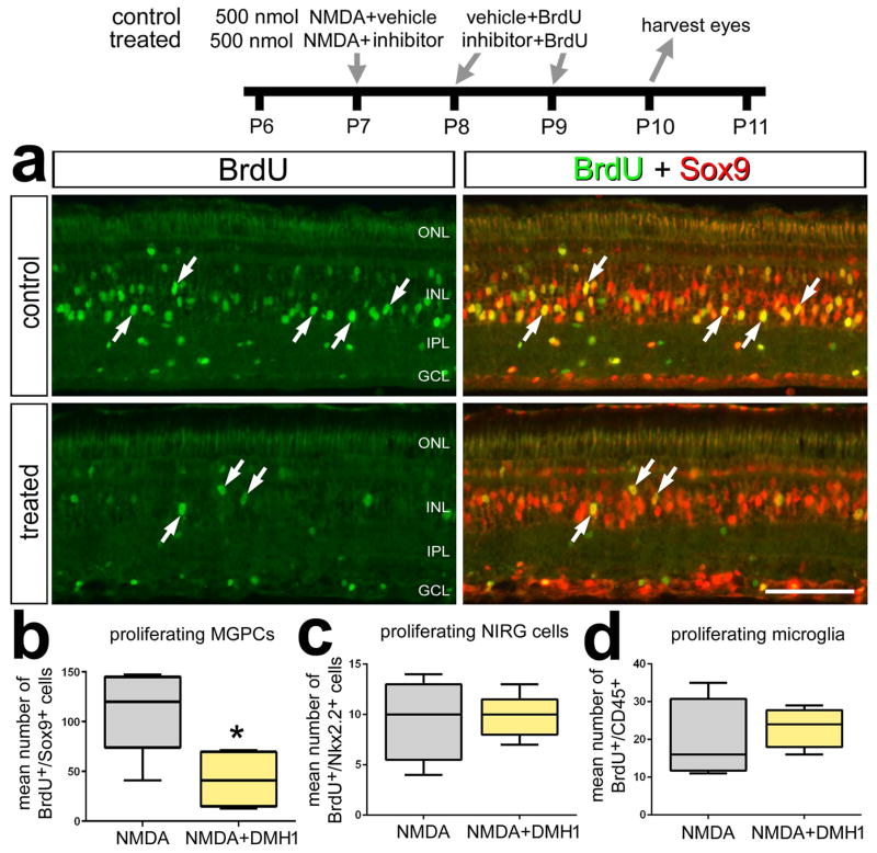
Figure 2.
Inhibition of BMP-signaling suppresses the formation of proliferating MGPCs in damaged retinas. Eyes were injected with 500 nmol NMDA+vehicle (control) or NMDA+DMH1 (treated) at P7, vehicle+BrdU or DMH1+BrdU at P8 and P9, and retinas were harvested at P10. Retinal sections were labeled with antibodies to BrdU (green) and Sox9 (red). The box plots illustrate the mean, upper extreme, lower extreme, upper quartile and lower quartile. Arrows indicate the nuclei of MGPCs and the calibration bar in panel a represents 50 μm. Significance of difference (*p<0.05, ***p<0.001) was determined by using a two-tailed paired student’s t-test. Abbreviations: INL – inner nuclear layer, IPL – inner plexiform layer, GCL – ganglion cell layer, ONL – outer nuclear layer.

