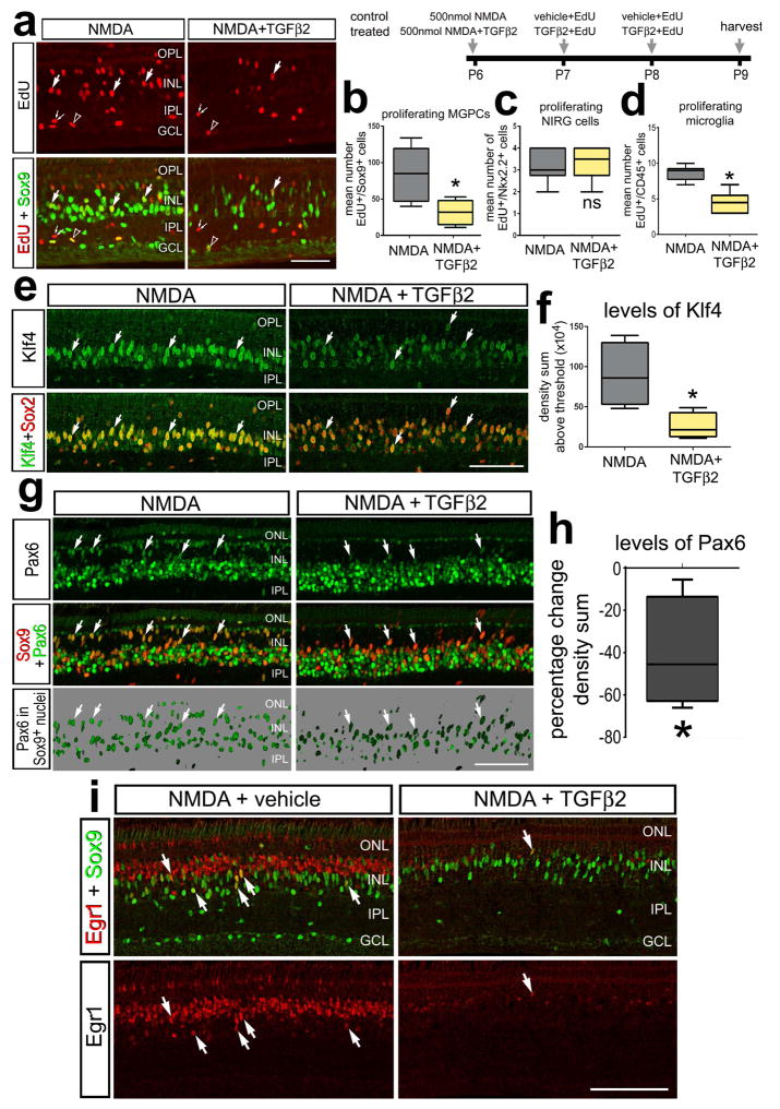
Figure 3.
TGFβ2 inhibits the formation of proliferating MGPCs in NMDA-damaged retinas. Eyes were injected with 500 nmol NMDA+vehicle (control) or NMDA+TGFβ2 (treated) at P6, vehicle+EdU or TGFβ2+EdU at P7 and P8, and retinas harvested at P9. Sections of the retina were labeled for EdU (red) and antibodies to Sox9 (green; a), Klf4 (green) and Sox2 (red; e), Pax6 (green) and Sox9 (red; g), or Sox9 (green) and Egr1 (red; i). The box plots illustrate the mean, upper extreme, lower extreme, upper quartile and lower quartile. The plots illustrate the number of proliferating MGPCs (b), NIRG cells (c), and microglia (d), levels of Klf4 in the nuclei of Müller glia/MGPCs (f), and percent change in the nuclear levels of Pax6 in Müller glia/MGPCs (h). Significance of difference (*p<0.05) was determined by using a two-tailed paired student’s t-test. Arrows indicate the nuclei of MGPCs. The calibration bar in each panel represents 50 μm. Abbreviations: INL – inner nuclear layer, IPL – inner plexiform layer, GCL – ganglion cell layer, ONL – outer nuclear layer.

