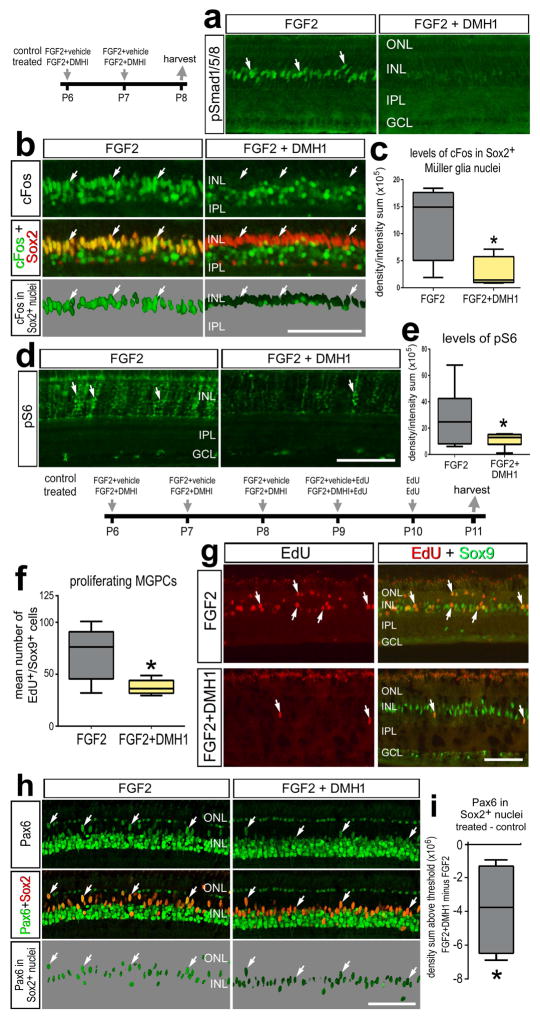
Figure 5.
Smad1/5/8-signaling is part of the cell-signaling network initiated by FGF2/MAPK that stimulates the proliferation of MGPCs in FGF2-treated retinas. a–e; Eyes were injected with FGF2+vehicle (control) or FGF2+DMH1 (treated) at P6 and P7, and retinas harvested at P8. f–i; eyes were injected daily with FGF2+vehicle (control) or FGF2+DMH1 (treated) at P6-P9, EdU alone at P10, and retinas harvested at P11. Sections of the retina were labeled with antibodies to pSmad1/5/8 (a), cFos (green) and Sox2 (red; b), or pS6 (d). The panels with a 70% grayscale background display cFos or Pax6 localized within the Sox9+ or Sox2+ nuclei of Müller glia (b), or Pax6 localized within the Sox2+ nuclei of Müller glia (h). The box plots illustrate the mean, upper extreme, lower extreme, upper quartile and lower quartile. The plots illustrates the control and treated levels (intensity sum) of cFos (c), levels of pS6 (e), or Pax6 (i) in Müller glia/MGPCs. The plot in f illustrates the abundance of proliferating MGPCs in control and treated retinas. Significance of difference (*p<0.05) was determined by using a two-tailed paired student’s t-test. Arrows indicate the nuclei of MGPCs. The calibration bar (50 μm) in panel b applies to a and b, the bar in d applies to d alone, the bar in g applies to g alone, and the bar in h applies to h alone. Abbreviations: INL – inner nuclear layer, IPL – inner plexiform layer, GCL – ganglion cell layer, ONL – outer nuclear layer.

