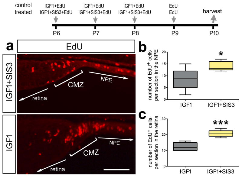
Figure 7.
Inhibition of Smad3 stimulates the proliferation of progenitors at the retinal margin. Eyes were injected with IGF1 (control) or IGF1+SIS3 (Smad3-inhibitor; treated) at P6, P7, P8, EdU alone at P9 and retinas were harvested at P10. Sections of the retina were labeled for EdU (a). The box plots illustrate the mean, upper extreme, lower extreme, upper quartile and lower quartile. The plots represent the numbers of proliferating cells in the non pigmented epithelium (200 μm anterior to the peripheral edge of the retina; b) and retinal margin (CMZ + retina; c). Significance of difference (*p<0.05, ***p<0.001) was determined by using a two-tailed paired student’s t-test. The calibration bar in panel a represents 50 μm. Abbreviations: CMZ – ciliary marginal zone, NPE - Non-pigmented epithelium.

