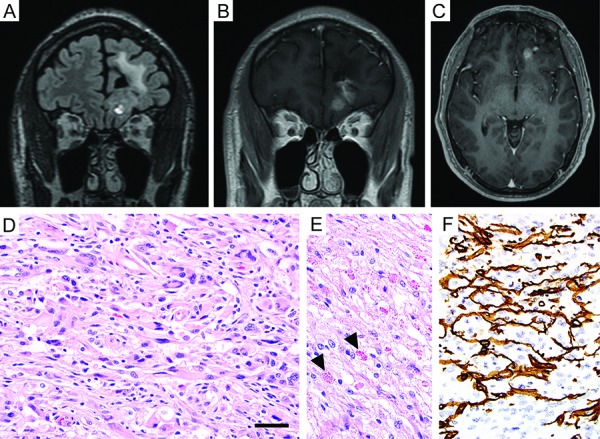
Figure 2. Imaging and histologic features of the pleomorphic xanthoastrocytoma resected from the left frontal lobe. A, B, C: Surveillance MR imaging 8 years after initial resection of a low-grade astrocytoma from the left frontal lobe demonstrated interval development of a new 7-mm focus of nodular enhancement adjacent to the prior resection cavity within the left medial orbital gyrus. Coronal T2/FLAIR-weighted image (A), coronal T1-weighted post-gadolinium image (B), and axial T1-weighted post-gadolinium image (C). D, E: Hematoxylin and eosin stained sections showing an astrocytic neoplasm with marked nuclear pleomorphism, numerous eosinophilic granular bodies (arrowheads), and occasional xanthomatous tumor cells with foamy cytoplasm. F: Immunostain for type IV collagen demonstrating abundant intercellular collagen deposition amongst the neoplastic astrocytes. Scale bar, 40 μm.

