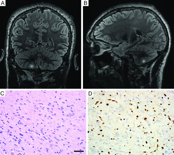
Figure 3. Imaging and histologic features of the diffuse astrocytoma resected from the right cerebellum. A, B: T2/FLAIR-weighted MR images demonstrating a hyperintense mass lesion in the right cerebellar hemisphere (coronal, A; sagittal, B). C: Hematoxylin and eosin stained section showing a diffuse glial neoplasm composed of neoplastic fibrillary astrocytes with elongate and irregular, hyperchromatic nuclei infiltrating through the cerebellar subcortical white matter. D: Immunohistochemistry for ATRX protein demonstrating absence of staining in the tumor cells with intact staining in entrapped non-neoplastic neurons and endothelial cells, consistent with somatic ATRX loss. Scale bar, 40 µm.

